

Disclaimer:
This is a popular science article, which aims to popularize the relevant knowledge in the field and promote science communication.
The video, pictures, text and other materials used in this article, if involved in the copyright of works, please contact us in a timely manner, we will properly handle according to law.
In addition, if there are errors in the content of the article, or inconsistent with the original journal article point of view, conclusion, also welcome to inform us, we will be the first time to modify, delete and so on.
At the same time, we warmly welcome scientific researchers and science enthusiasts in related fields to contribute, recommend, or discuss cooperation to jointly promote the development of industrialization.
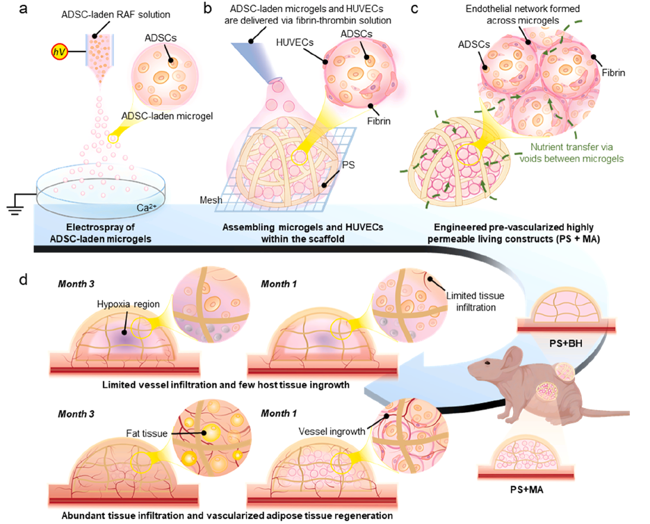
Breast deformity following mastectomy represents a prevalent clinical challenge in soft tissue reconstruction. Current strategies combining 3D-printed polymeric scaffolds with hydrogels are capable of loading stem cells and exhibit promising regenerative potential. Nevertheless, when applied to repair large-volume defects, such constructs frequently suffer from insufficient interconnected internal micropores. Oxygen and nutrients cannot diffuse efficiently deep into the implant, resulting in persistent localized hypoxia within the scaffold. This markedly impedes the ingrowth of neovessels and limits the regeneration of large-volume adipose tissue. Recently, the research team led by Professor He Jiankang and Dr. Meng Zijie at Xi’an Jiaotong University proposed an innovative assembly strategy of scaffolds integrated with cell-laden microgels. This approach simultaneously addresses three core bottlenecks: mechanical support, oxygen and nutrient supply, and prevascularization. Encouraging preliminary outcomes have been achieved, offering an innovative modular solution for generating large-volume vascularized adipose tissue.
The related study entitled Scaffold-assisted assembly of cell-laden microgels to engineer permeable living constructs for vascularized adipose tissue regeneration has been published in Acta Biomaterialia.
Highlights: Innovation and Breakthrough
- Binary composite structural innovationThe integration of stem cell-laden microgels with 3D-printed PCL scaffolds overcomes two prominent bottlenecks in the industry: poor oxygen supply in bulk hydrogels and insufficient cell retention in bare scaffolds.
- In-situ prevascularized microenvironmentNatural intergranular channels among microgels accommodate endothelial cells, facilitating the in vitro fabrication of trans-particle endothelial networks and enhancing adipogenic differentiation of stem cells.
- Volume-stable regenerative systemScaffolds provide mechanical support to prevent implant collapse, while interconnected pores within microgels improve oxygen transport and substantially mitigate hypoxia in defective regions.
- Precisely tunable particle sizeElectrospraying enables microgel size regulation ranging from 200 μm to 600 μm. Microgels of 200 μm are validated to achieve optimal comprehensive performance in mass transport and cell viability.
WHAT: Research Content
In this study, RGD-grafted alginate-fibrin (RAF) electrosprayed microgels were adopted as carriers for adipose-derived stem cells (ADSCs). Combined with 3D-printed helical PCL scaffolds (PS), composite living constructs designated PS+MA were fabricated. Comprehensive multi-dimensional characterizations were carried out as follows:
- Electrospray parameters were optimized to produce microgels of varying sizes, followed by characterization of swelling behavior, degradation profiles and stem cell viability.
- Microgels were assembled with scaffolds. μCT scanning and fluid diffusion simulation were performed to characterize pore architecture and permeability.
- In vitro comparisons were conducted with the bulk hydrogel group (PS+BH) to evaluate cell survival and proliferation. Human umbilical vein endothelial cells (HUVECs) were introduced to construct prevascularized structures, and adipogenic differentiation levels were quantified.
- COMSOL finite element simulation was employed to model oxygen distribution and quantify hypoxic discrepancies between groups.
- Subcutaneous implantation in nude mice was performed. At week 4 and week 12, magnetic resonance imaging (MRI), angiography and immunostaining were utilized to assess angiogenesis, adipose regeneration and hypoxia alleviation.
WHAT: Research Background and Significance
Post-mastectomy breast defects not only disfigure patients’ breasts but also impose severe psychological burdens. 3D-printed scaffolds combined with adipose tissue engineering provide a viable strategy for repairing such defects, yet two major technical hurdles remain unresolved. Firstly, standalone 3D-printed scaffolds fail to form a favorable microenvironment for cell colonization; most cells merely adhere to the scaffold surface, leading to heterogeneous cell distribution. Secondly, filling the scaffold bulk with monolithic hydrogel blocks pore channels. The loss of interconnected pores restricts oxygen and nutrient diffusion, triggering massive hypoxic cell necrosis in the interior. In addition, host neovessels struggle to infiltrate deep into the biomaterial, ultimately resulting in low efficiency of adipose regeneration. Meanwhile, granular microgels alone possess poor mechanical properties and cannot sustain the spatial volume required for tissue regeneration in vivo. Accordingly, there is an urgent clinical demand for novel tissue-engineered constructs that integrate mechanical stability, high mass transport permeability, uniform cell seeding capacity, and the capability to support prevascularization.
The Solution: the technologicaledge
1. Overall fabrication workflow
Microscale fibrous porous frameworks are fabricated via electrohydrodynamic forming technology. Polydopamine-mediated interfacial conjugation enables drug loading, breaking the technical barrier that thermosensitive bioactive agents cannot be incorporated into melt-printed scaffolds. The system exhibits sustained, staged drug release profiles matching medication requirements in the early stage of bone defect repair. Leveraging surface modification, the scaffolds combine load-bearing mechanical properties with favorable biological interfaces. Multiple cellular safety assessments confirm no cytotoxicity at designated drug dosages. Local targeted delivery fundamentally avoids adverse systemic reactions induced by systemic medication.
2. Intergranular interconnected pores enhance mass transport and eliminate deep-layer hypoxia
Microgel assemblies with a particle size of 200 μm achieve a void fraction of up to 41% with interconnected pores. Fluid infiltration tests and finite element simulations verify the hierarchy of mass transport efficiency: MA200 > MA400 > MA600 > bulk hydrogel (BH). Severe diffusion resistance occurs within bulk hydrogels, generating extensive central hypoxic regions. Interstitial gaps between microgels form rapid exchange pathways for nutrients, oxygen and metabolic wastes, homogenizing oxygen distribution inside constructs and markedly shrinking hypoxic areas.
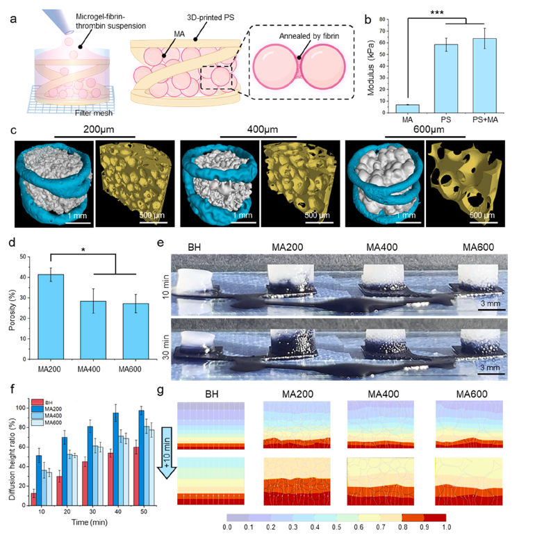
Figure 2 Pore architecture, dye penetration and substance diffusion simulation results of microgel assemblies with uniform particle size
3. Synergistic mechanics via rigid-soft stratification balancing shaping capacity and biological activity
Implantation experiments in critical-sized rat calvarial defects demonstrate that drug-loaded scaffolds effectively modulate macrophage polarization at wound sites, suppress persistent inflammatory responses and establish a favorable microenvironment for tissue repair. Co-culture systems verify positive crosstalk between immune cells and osteoblasts. In vivo detection confirms significantly improved volume of newly formed bone and superior trabecular quality compared with control groups. Relying on direct osteogenic induction and immune regulation, efficient bone defect repair is realized.PCL scaffolds undertake primary mechanical support. The compressive modulus of bare PCL scaffolds reaches 59 kPa, rising to 64 kPa in composite PS+MA constructs, whereas pure microgel assemblies (MA) only exhibit a modulus of 6.9 kPa with insufficient mechanical rigidity. The composite system realizes a functional combination: rigid scaffolds lock regenerative contour, while soft microgels provide biomimetic cellular microenvironments to prevent collapse and shrinkage post-implantation.
4. Interstitial channels accommodate endothelial cells for in vitro prevascularization to boost adipogenesis
Intergranular voids serve as independent cell niches. HUVECs spread along particle surfaces and form continuous endothelial networks spanning multiple microgels after 14 days of culture. Paracrine signals released by endothelial cells upregulate adipogenic genes in ADSCs (PPARγ, FAS, adiponectin), yielding substantially larger lipid droplets compared with non-vascularized controls. In vivo mechanism: prefabricated human endothelial networks act as angiogenic templates to recruit and fuse with host endothelial cells, accelerating global angiogenesis.
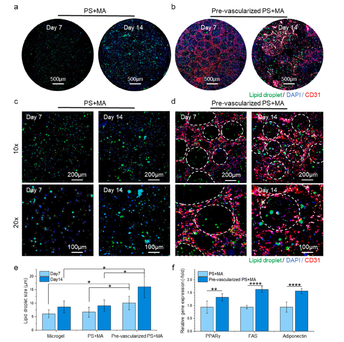
Figure 3 Fluorescent staining of endothelial networks and lipid droplet formation within prevascularized constructs
5. Homogenously distributed cells sustain long-term stem cell survival and proliferation
In the bulk hydrogel group, only surface-layer cells gain access to nutrients. Cell viability declines from 71% to 41% after 14 days of culture. Benefiting from efficient mass transport through interstitial channels, the PS+MA group maintains cell viability at 78% at day 14, with a Ki67-positive proliferative cell ratio over twice that of the control group. This innovation fundamentally resolves the problem of central cell necrosis in thick hydrogel constructs.
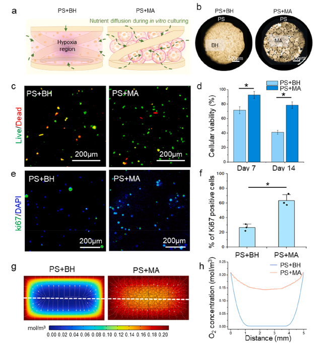
Figure 4 Comparison of cell survival, proliferation and oxygen distribution simulation between bulk hydrogels and microgel composite constructs
6. Concurrent improvement of vascular infiltration, hypoxia relief and adipose regeneration in vivo
Vascular ingrowth: At 12 weeks post-implantation, prevascularized PS+MA groups display markedly higher microvessel density than the PS+BH group; host tissue infiltrates deep into constructs along microgel gaps.Hypoxia mitigation: The positive area of hypoxia marker HIF-1α is significantly lower than that in bulk hydrogel groups.Stable adipose regeneration: Quantitative MRI at week 12 reveals an adipose tissue ratio of 39% in the PS+MA group, outperforming bare scaffolds and bulk hydrogel-filled groups. Lipid droplet protein staining further confirms the optimal maturity of regenerated adipose tissue.
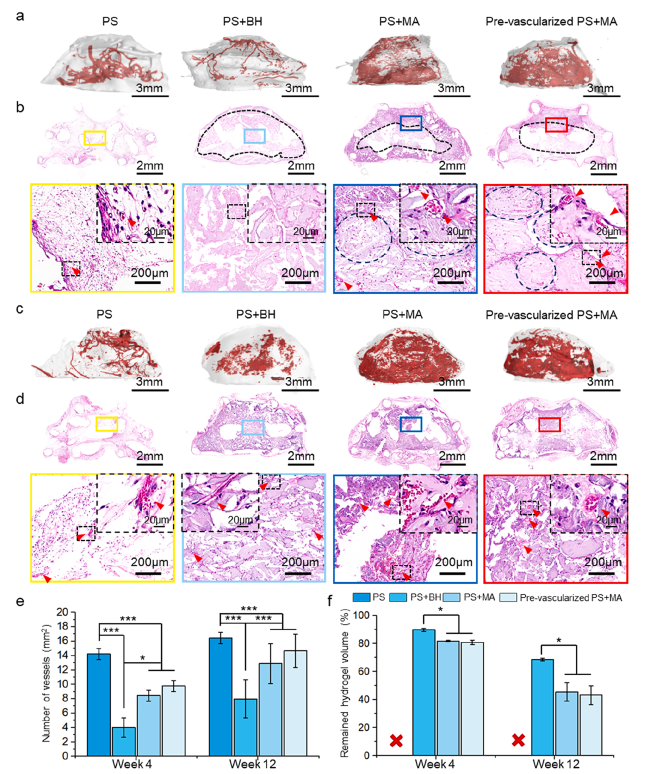
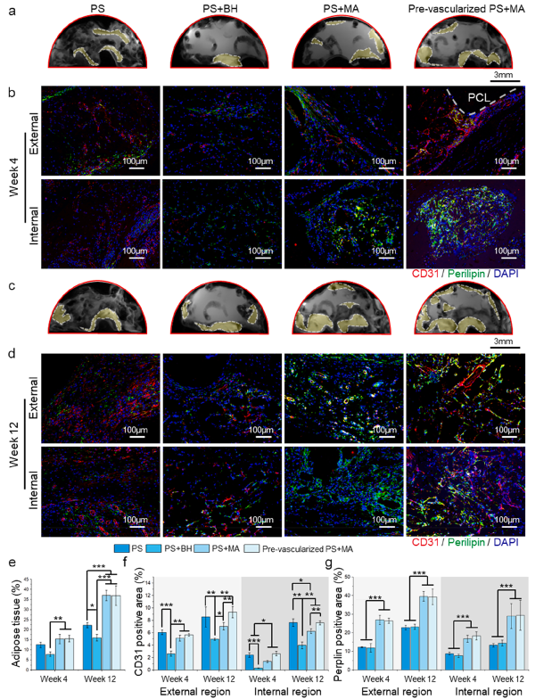
Figure 5 In vivo microangiography and MRI quantitative detection of adipose regeneration
Conclusions
The scaffold-assisted microgel assembly strategy proposed in this study ingeniously integrates the mechanical superiority of 3D-printed scaffolds and the high permeability of microgel assemblies. Its core value lies in the adoption of microscale channel design, which fundamentally addresses the long-standing limitation of conventional hydrogels — the dilemma of being able to encapsulate cells yet failing to sustain cell viability. This work establishes a novel construct paradigm featuring structural stability and efficient mass exchange for vascularized soft tissue regeneration. The strategy holds great promise for translation to other soft tissue repair scenarios demanding rapid vascularization.