

Disclaimer:
This is a popular science article, which aims to popularize the relevant knowledge in the field and promote science communication.
The video, pictures, text and other materials used in this article, if involved in the copyright of works, please contact us in a timely manner, we will properly handle according to law.
In addition, if there are errors in the content of the article, or inconsistent with the original journal article point of view, conclusion, also welcome to inform us, we will be the first time to modify, delete and so on.
At the same time, we warmly welcome scientific researchers and science enthusiasts in related fields to contribute, recommend, or discuss cooperation to jointly promote the development of industrialization.
The corresponding work entitled Strain-Driven Topological Reorganization in Soft Fibrin Nanofibrous Networks Enabling Tissue-Like Alignment has been published in Advanced Materials.
Core Innovations & Breakthroughs
- Innovative fabrication methodologyFor the first time, uniaxial strain is applied during the dynamic fibrillogenic window of fibrin gel; permanent ordered architectures are locked in place via progressive fibrin crosslinking. The team identified a 90-second pre-gelation optimal processing window and a saturation threshold for fibre alignment at 1.6-fold stretch. This template-free, external-field-free technique permits scalable manufacturing of 3D aligned cell-laden matrices.
- Quantitative geometric theory for pore straighteningA geometric model of pore straightening explains the intrinsic mechanism governing alignment saturation at 1.6-fold stretch from a microscale topological perspective. Combined with rheological characterisation and finite element modelling, the study delineates the mechanical mechanisms through which crosslink density regulates fibre mobility and force transmission balance within the network. These findings deliver quantitative design guidelines for controlling alignment in fibrous biological hydrogels.
- Integrated structural and functional engineeringOptimisation of scaffold lattice geometries yields biomimetic layered myocardium. Integration with piezoelectric scaffolds enables endogenous electrical stimulation triggered by cellular contraction, establishing an integrated system of “biomimetic layered myocardium + piezoelectric self-stimulation”. The platform concurrently enhances cardiomyocyte structural maturation and electrophysiological function for cardiac repair applications, with broad applicability across multiple soft tissue engineering modalities.
WHAT:Research Scope
Conventional strategies for generating aligned 3D cell-laden fibrous matrices depend on templates or external physical fields, lack quantitative mechanical regulation, and struggle to simultaneously recapitulate layered biomimetic myocardial architecture while tuning electrophysiological function. To address these bottlenecks, the authors present a gelation-coupled strain-induced alignment strategy. By modulating pre-gelation duration, stretch ratio and scaffold geometry, stable ordered fibrin networks are fabricated. A geometric model rationalises the 1.6-fold stretch alignment saturation phenomenon. Optimised lattice designs enable production of centimetre-scale uniaxially aligned and multi-layer biomimetic myocardial constructs, whose translational utility in engineered cardiac tissue and myocardial repair is validated.
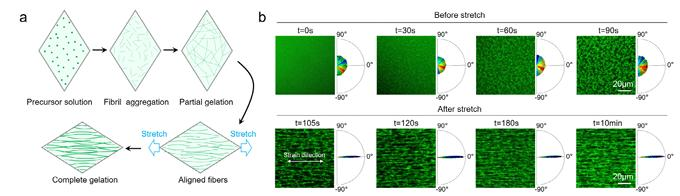
Figure 1: Schematic illustration of the overall design
Technical Advantages & Solutions
1. Development of a tunable viscoelastic culture platform
An electrohydrodynamically printed diamond-patterned PCL scaffold serves as the substrate for strain assembly. Fibre organisation and post-unloading recoil behaviour are compared across three pre-gelation timepoints (60 s, 90 s, 10 min). Rheological testing quantifies differences in storage modulus across distinct crosslinking stages, confirming that the intermediate 90 s crosslinking window strikes a favourable balance between fibre slippage and bulk load-bearing capacity. Uniaxial stretch is applied over a gradient of 20%–80% deformation; fibre/cell alignment indices and pore aspect ratios are quantified systematically. A polygon pore straightening geometric model elucidates the microtopological basis for alignment saturation at 1.6-fold stretch. Finite element simulations further reveal disparities in stress propagation and network rearrangement under weak, intermediate and fully crosslinked conditions.
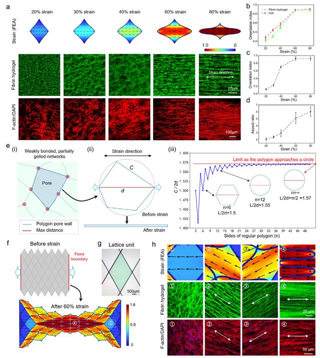
Figure 2: Strain-dependent saturation of fibrin and cellular alignment from unit elements to scaffold arrays
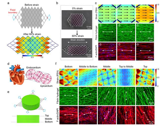
3. Biological validation of cardiomyocyte structure and contractile function within aligned matrices
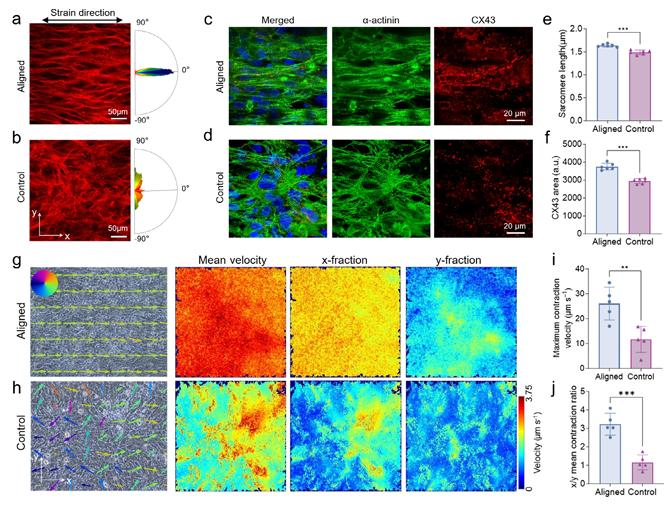
Neonatal rat cardiomyocytes are encapsulated within strain-aligned fibrin matrices, with non-aligned disordered hydrogels as controls. Immunofluorescent analysis of sarcomere length and connexin 43 (CX43) gap junction expression verifies that aligned microenvironments promote cardiomyocyte structural maturation. High-speed imaging paired with optical flow tracking algorithms quantifies spontaneous contractile dynamics. Comparisons of peak contraction velocity and directional contractile partitioning along x/y axes confirm that aligned substrates drive rapid, synchronous, anisotropic directional contraction in engineered cardiac tissue.
4. Development and multifunctional validation of piezoelectric composite functional scaffolds
Multi-material electrohydrodynamic co-printing is utilised to manufacture PCL/PVDF piezoelectric composite scaffolds. Mechanical properties, PVDF electroactive phase transformation and electrical signal output are characterised. Results confirm that incorporation of piezoelectric components does not disrupt strain-induced fibre alignment. Electropacing protocols and calcium transient kinetic analysis are performed to compare pacing thresholds, maximum synchronous capture frequency and calcium recovery rates between pure PCL and piezoelectric composite groups. The data verify that spontaneous cardiomyocyte contraction activates endogenous piezoelectric stimulation, comprehensively enhancing cardiac electrophysiological performance. Finally, the full fabrication workflow is validated for building in vitro cardiac models and implantable patches for myocardial infarction repair. The framework is readily adaptable for constructing diverse anisotropic soft tissues including skeletal muscle and peripheral nerve.
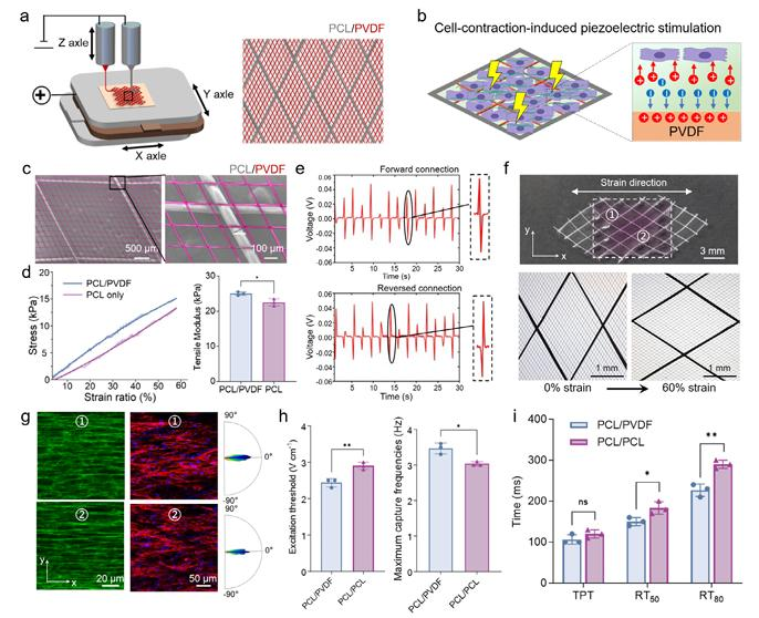
Figure 5: Electrohydrodynamically printed piezoelectric diamond scaffolds enhance electrophysiology of aligned cardiac tissue
Conclusions
This study establishes a gelation-coupled strain locking strategy. Uniaxial stretching is applied within the fibrin hydrogel gelation window, and the equilibrium between fibre slippage and network crosslinking generates permanently retained anisotropic architectures. The geometric model clarifies the mechanism of alignment saturation at 1.6-fold stretch. Optimised scaffold lattices enable scalable fabrication of large-area uniaxially aligned and transmural layered biomimetic myocardial matrices. Aligned microenvironments significantly boost cardiomyocyte structural maturation and directional contraction. When combined with PCL/PVDF piezoelectric scaffolds, spontaneous cellular contraction generates intrinsic electrical stimulation to further refine cardiac electrophysiology and calcium handling. The technique features straightforward operation and excellent cytocompatibility, suitable for manufacturing in vitro cardiac models and implantable constructs for myocardial repair. Moreover, this platform can be translated to a broad spectrum of anisotropic soft tissue engineering systems encompassing skeletal muscle and peripheral nerve regeneration.